Tirouvanziam Lab Awarded ICI Mini-Pilot
Graduate student Camilla Margaroli, MSc and PI Rabin Tirouvanziam, PhD were recently awaded an ICI Core mini-pilot: "Next Level of Fluorescence." Read below about the status of their project.
High Resolution Imaging of PD1 Signaling in Airway Cells
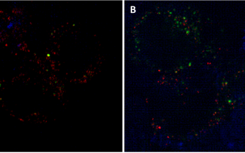
The main cause of morbidity and mortality in cystic fibrosis is lung disease, which is characterized by sustained neutrophil recruitment from blood to the airway lumen. Recent data from our group suggest that neutrophil recruitment correlates with exhaustion of resident airway macrophage, as evidenced by expression of the regulatory receptor PD1. To gain further insight into this mechanism, we collected airway leukocytes from CF infants by bronchoalveolar, then stained and imaged these cells on high-resolution microscopes at the Integrated Cellular Imaging Core. First, we used the Olympus FV1000 confocal microscope to assess optimal staining. Second, we used the GE Delta Vision OMX Blaze microscope with SIM (Structured Illuminated Microscopy) to determine colocalization of membrane PD1 (green) with its intracellular protein phosphatase partner SHP2 (blue), as well as lipid raft signaling platforms (red) to assess pathway activation. Images shown below illustrate this set of experiments in CF airway macrophages and neutrophils.
Figure: PD1 expression in CF airway macrophages and neutrophils. Airway cells from bronchoalveolar lavage of CF patients were fixed, stained and imaged on the GE Delta Vision OMX Blaze microscope using SIM. Macrophages (A) and neutrophils (B) were assessed for colocalization of PD1 (green), lipid rafts signaling platforms (red) and intracellular mediator SHP2 (blue).